When you notice heavy leg swelling, tight cramping, or unusual pain, we need a clear picture of your circulation to find out what is wrong. Doppler ultrasound is a noninvasive test [1] that measures the direction and speed of blood cells as they move through vessels [2].
We use this test every day in the clinic because it gives direct answers without surgery or discomfort. When you understand the process, the anxiety of the unknown drops. Understanding Vascular Doppler Ultrasound: What It Shows & How to Prepare helps you feel confident and ready for your visit.
Here is exactly what you will learn in this guide:
- How the test checks your veins and arteries safely.
- What a clot or poor blood flow looks like on our screen.
- Why a surgeon-performed ultrasound matters for your treatment.
- What you need to do before you walk into the clinic.
Table of Contents
What is a vascular Doppler ultrasound and what does it show?
We rely heavily on this imaging tool because ultrasound does not use ionizing radiation [2]. It is entirely safe, even if we need to repeat the scan multiple times to monitor your progress. The machine uses high-frequency sound waves to create live pictures of your blood vessels.
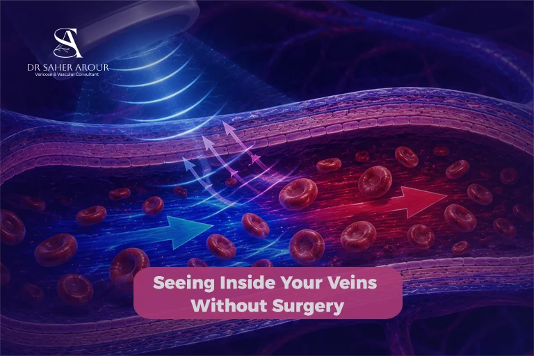
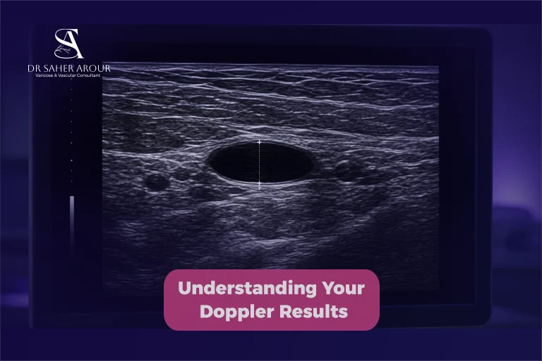
A standard B-mode ultrasound veins scan only shows the static walls. To understand your symptoms, we need to see movement. When I look at the screen, colour Doppler blood flow shows me exactly which way your blood is traveling. If there is a sudden block or blood flowing backwards, the Doppler highlights it immediately. We get a real-time, moving map of your circulation.
What Conditions a Vascular Doppler Can Detect
We use this scan to evaluate two main systems: your veins, which carry blood back to your heart, and your arteries, which deliver oxygen-rich blood to your body.
Venous Conditions: DVT, Venous Reflux, Varicose Veins
When blood struggles to move up your legs, it pools in the lower veins, causing heaviness and swelling. I see how frustrating and uncomfortable this can be for patients who spend all day on their feet. Through a dedicated duplex ultrasound veins evaluation, we look for:
- Deep Vein Thrombosis (DVT): Compression ultrasonography is the primary imaging modality for diagnosing DVT [3]. Ultrasound has high sensitivity and specificity for proximal DVT [3]. Accurate DVT ultrasound diagnosis is critical for your safety.
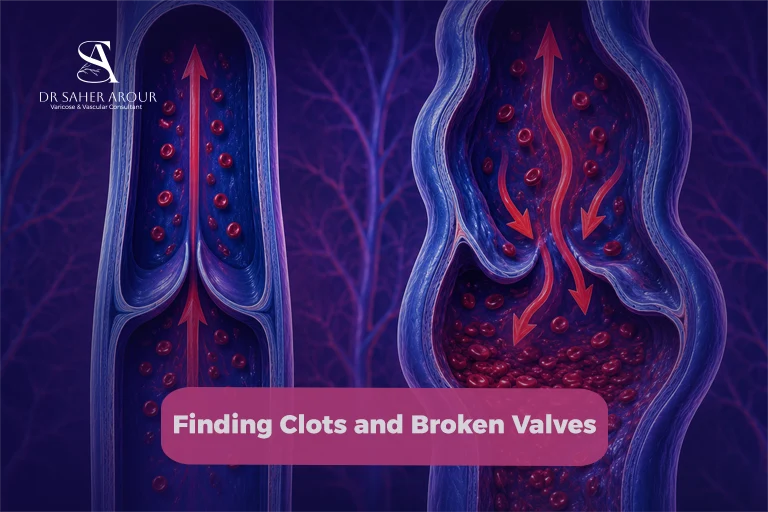
- Venous Reflux: A venous reflux ultrasound checks if tiny valves inside your veins are failing and letting blood fall backward.
- Varicose Veins: Backward flow causes veins to stretch and bulge. The Doppler pinpoints exactly where these broken valves are located.
Arterial Conditions: Stenosis, Occlusion, Aneurysm
In your arteries, cholesterol and plaque can slowly build up over the years. This narrows the vessel, a condition we medically call stenosis. If you picture a kink in a garden hose making the water spray faster, it is a very similar concept. If plaque completely blocks flow, it causes an occlusion, leading to severe pain when walking. A Doppler scan arteries test measures how fast blood squeezes past these narrowings.
We also check for aneurysms, which are weak spots in the artery wall that balloon outward. By measuring the artery precisely, we can plan the safest treatment well before it becomes a medical emergency.
How the Examination Works: What Happens During the Scan
If you are feeling nervous, take a deep breath. Ultrasound exams are painless [2]. There is no enclosed tube, no loud noises, and no needles.

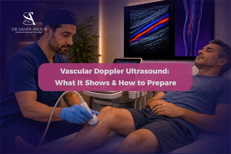
You will lie comfortably on an examination table. We apply a clear, warm gel to your skin to help the sound waves travel efficiently. Next, we slide a small handheld probe over your skin. You may feel a firm pressure as we press down to see deeper vessels clearly, but it is not painful.
Occasionally, I might ask you to take a deep breath or bear down gently. The exam usually takes less than an hour [2]. You can just lie back and let us do the work. Once we finish, you can step right off the table and carry on with your day.
Why a Surgeon-Performed Ultrasound May Differ From a Radiologist’s Report
A hospital imaging center provides a very helpful general report. However, interpretation varies based on operator experience. When I perform the scan personally in the clinic, I am actively mapping out your specific treatment plan.
| Evaluation Aspect | Standard Radiology Scan | Surgeon-Performed Clinic Scan |
| Primary Goal | To write a general diagnostic report. | To diagnose and visually map the exact surgical fix. |
| Real-Time Assessment | The referring doctor reads static images later. | The treating doctor evaluates the live flow directly. |
| Patient Connection | Often performed by a separate technician. | I connect your physical symptoms to the screen instantly. |
This hands-on approach lets me trace abnormal, twisting veins precisely. We see the problem through a surgeon’s eyes, saving you time and giving you a highly tailored evaluation.
What the Doppler Report Means: Decoding the Key Terms
After the scan, your report might contain unfamiliar clinical words. My goal is to make sure you understand exactly what they mean for your health.
Reflux Duration: What a Positive Reflux Finding Means
You will likely see notes about reflux duration. Reflux simply means blood is flowing in the wrong direction. The Doppler measures exactly how long that backward flow lasts. If the blood falls backward for longer than an accepted threshold, we consider it a positive reflux finding. We use this to confirm your vein valves need medical support.
Vein Diameter: What Is Significant
The report lists the diameter of your veins in millimeters. When veins hold pooled blood, they stretch and grow wider than they should. While there is no single universal number for everyone, a significantly wider vein usually points to unhealthy pressure building up inside. I look at this width alongside your pain to decide the best step forward.
How to Prepare for a Vascular Doppler Scan
I want to make your clinic visit as smooth as possible. Proper vascular ultrasound preparation is simple:
- Diet and Medication: No special preparation is required for most Doppler ultrasound exams [2]. You do not need to pause your medications, and you can eat and drink normally.
- Clothing: Wear loose, comfortable clothing. We might ask you to change into a simple clinic gown so we can reach your leg or arm easily.
- Abdominal Scans: If we are examining blood vessels deep in your abdomen, we might ask you to fast for a short time beforehand, but my team will always give you clear instructions.
Frequently Asked Questions
Is a vascular Doppler ultrasound painful?
No. Doppler ultrasound is a noninvasive test [1] and ultrasound exams are painless [2]. You may feel slight pressure from the probe, but the test does not involve needles, incisions, or radiation exposure.
How long does a vascular ultrasound take?
The exam usually takes less than an hour [2]. The exact time depends on the specific area of your body being examined and the complexity of your circulation.
Can a Doppler ultrasound miss a DVT?
Doppler ultrasound is highly accurate, showing high sensitivity and specificity for proximal DVT in the larger upper leg veins [3]. However, in rare cases involving very early-stage clots or tiny calf veins, additional imaging tests might be required.
Do I need to fast before a vascular ultrasound?
No special preparation is required for most Doppler ultrasound exams [2]. Fasting is usually not needed for arm or leg scans. However, specific abdominal scans may require you to fast.