
How to read a vascular ultrasound report and see the medical jargon, especially during pregnancy when your body is already going through so many changes, it’s normal to feel a bit lost among all those numbers. I’m here to help. Let’s decode these measurements, numbers, and classifications together.
Educational Disclaimer: This content is for informational purposes only and is not a substitute for professional medical advice, diagnosis, or treatment.
We won’t focus on how the scan is done today, but rather on helping you read the actual document by yourself, so you can:
- Identify exactly which blood vessel was examined.
- Recognize standard disease severity classifications.
- Make sense of objective measurements like blood flow velocities.
- Know exactly when a finding needs urgent attention.
Keep reading the article and bookmark this information to easily refer back to it later.
Table of Contents
How to Read a Vascular Ultrasound Report?
To understand your vascular ultrasound report, start by finding the examined blood vessels, checking the blood flow speed measurements, and reading the doctor’s final impression for any abnormalities.
Always remember that a single number on that paper doesn’t give you the full picture. As specialists, we evaluate the severity, location, and extent of these findings [1]. Your physical symptoms—like leg swelling during pregnancy—and your medical history are what give this data real meaning. If you need a broader look at how we evaluate your circulation, feel free to visit our Vascular Health Pillar.
The Anatomy of a Vascular Ultrasound Report: Common Standard Sections
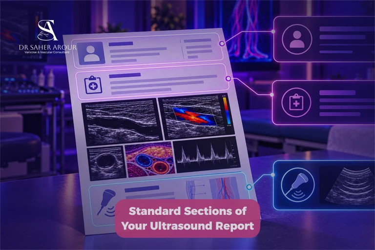
Every report follows a logical structure. Here is what expecting mothers and general patients will typically see as they read through the document:
Section 1: Patient Demographics and Indication for the Test
Your report starts with your basic details and the “indication.” This is simply the medical reason your doctor requested the test—such as “leg pain,” “pregnancy-related swelling,” or “stroke screening.” It gives the reviewing specialist the exact clinical context they need before looking at your Vascular Screening Tests.
Section 2: Technique and Type of Ultrasound Performed
This section tells us exactly how the technician performed the examination. For example, if you had your carotid artery checked, the report should specify that the assessment used gray-scale, color Doppler, and spectral Doppler ultrasound [2].
These imaging techniques work together so we can visualize the vessel structure and accurately measure the speed of your blood flow.
Three Categories of Key Terms You Will See: Decoded in Plain English
You don’t need a medical degree to understand your findings. Let’s break the most common jargon down into three simple categories so you know exactly what you are looking at.
Category 1: Flow Terms (Patent, Stenosis, Occlusion, Thrombus)
- Patent: Good news! The vessel is open and blood flows through it normally.
- Stenosis: The blood vessel is narrowed.
- Occlusion: The vessel is completely blocked with no detectable flow.
Category 2: Vein Valve and Reflux Terms (Competent, Reflux, Incompetent, Valsalva)
If you’re reading a venous report for your legs—which is a very common scan for pregnant women experiencing heavy or swollen legs—you might spot the word “reflux.” Venous reflux just means blood is flowing backward through a vein because the tiny valves inside aren’t closing tightly.
If your report says the valves are “competent,” that means they’re working perfectly to keep your blood moving in the right direction. For more details on this, you can check our Varicose Veins Diagnosis resources.
Category 3: Plaque and Wall Terms (Intima-Media Thickness, Plaque, Calcification)
Plaque is a very common finding that simply means there is a buildup of fat or cholesterol on the artery wall. Instead of getting lost in the exact millimeters on your report, here is what doctors look for to officially classify it as plaque [3]:
- A buildup pushing into the blood flow space by at least 0.5 mm.
- An overall thickness greater than 1.5 mm.
You might also see IMT (Intima-Media Thickness) measurements. Don’t worry if your IMT isn’t compared to a strict “normal” number. We avoid using a single cutoff because your artery’s normal thickness naturally changes depending on your age, sex, and race [4].
Common Findings by Test Type: What Each Report Is Looking For
Depending on which blood vessels were checked, your ultrasound report will focus on specific measurements.
Carotid Duplex: Stenosis Grading (50%, 70%, Near-Occlusion)
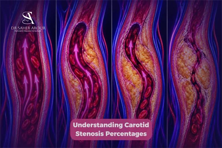
While checking carotid arteries is less tied to pregnancy, if you’re looking at a Carotid Artery Disease report, it uses standard categories to grade any narrowing [2]. We determine these grades mostly by checking the Peak Systolic Velocity (PSV)—which is simply how fast your blood is moving—and whether plaque is visible.
Let’s say your artery is like a garden hose. If you pinch the hose, the water sprays faster. Your arteries work exactly the same way.
By measuring how fast your blood moves (PSV), we can tell how narrow the artery is. Please don’t panic if you see high numbers; they are just a way for doctors to evaluate your circulation safely. Here is how we break them down:
- Normal: Blood speed (PSV) is under 180 cm/sec, and there’s no plaque [5].
- 50% to 69% Stenosis: Blood speed is between 180 and 230 cm/sec, with visible plaque [5].
- Greater than 70% Stenosis: Blood speed exceeds 230 cm/sec [5].
- Near Occlusion: Plaque is clearly visible, and blood flow is severely restricted, making the speed highly variable (it could be high, low, or even undetectable) [5].
- Total Occlusion: The artery is completely blocked with no detectable flow [5].
Please remember that simply finding plaque doesn’t automatically mean you have severe narrowing [6]. Its presence and structure just help us evaluate your cardiovascular health and overall risk to protect you moving forward [7].
Lower Extremity Venous Duplex: DVT and Reflux Findings
During pregnancy, the increased pressure on your pelvic veins makes checking your leg veins particularly important. The ultrasound targets two main issues: blockages (like a blood clot or DVT) and reflux. During the test, the technician gently compresses your veins. If a vein can’t be compressed, it usually points to a clot. When checking for reflux, they measure the exact duration of backward flow to see if your valves are struggling to push blood back up to your heart.
Arterial Duplex and ABI: Peripheral Artery Disease Assessment and Chronic Limb-Threatening Ischemia
For leg arteries, your report may include an Ankle-Brachial Index (ABI). I recommend a resting ABI to confirm a Peripheral Artery Disease diagnosis if your physical exam or medical history suggests it [8]. Looking at your ABI results? Here is exactly what those ranges mean for your leg circulation:
| ABI Category | Measurement Range | What It Means |
| Normal | 1.00 to 1.40 | Healthy arterial blood flow in your legs [8]. |
| Borderline | 0.91 to 0.99 | Flow is slightly reduced and requires monitoring [8]. |
| Abnormal | 0.90 or less | Indicates reduced blood flow, supporting a PAD diagnosis [8]. |
| Noncompressible | Greater than 1.40 | Your arteries are stiff (often due to calcification), meaning the ABI cannot be accurately read right now [8]. |
In cases of severe disease, you might spot the term Chronic Limb-Threatening Ischemia (CLTI). This is the updated medical term for what used to be called “critical limb ischemia.” Rather than just a confusing label, CLTI simply means your arterial blood flow is severely restricted, leading to specific issues [9]:
- Chronic pain in your legs or feet while resting (lasting more than two weeks).
- Wounds or ulcers that refuse to heal.
- Noticeable tissue damage or gangrene.
Red Flags That Require Urgent Follow-up With a Vascular Specialist
While only your doctor can give you definitive medical advice based on your full file, certain findings shouldn’t wait. For example, if your ABI falls to 0.40 or below, it points to severe disease [10].
Similarly, a finding of total carotid occlusion or a near-occlusion pattern with unstable plaque needs prompt review by a vascular surgeon. Don’t panic, but do schedule an appointment quickly so we can discuss your management plan and protect your health and your baby’s wellbeing if you are expecting.
FAQ Section: People Also Ask
What does “patent” mean on a vascular ultrasound report?
A patent vessel is simply open and allows blood to flow through it. However, the term doesn’t automatically mean the vessel is completely normal or healthy.
What is the difference between stenosis and occlusion?
Stenosis means the blood vessel is narrowed, while occlusion means the vessel is completely blocked with no detectable flow on the examination.
What does reflux in a vein mean?
Venous reflux means blood is flowing backward through your vein, usually because the small valves inside aren’t closing properly.
Should I be worried if my carotid ultrasound shows plaque?
Plaque indicates some level of carotid atherosclerosis, but you shouldn’t panic. Its clinical importance depends entirely on how much it narrows the artery and your overall clinical context.
What is a normal ABI value and what does it mean if it is low?
A resting ABI between 1.00 and 1.40 is completely normal. If it drops to 0.90 or below, it’s considered abnormal and strongly suggests peripheral artery disease.
References
- [1] https://intersocietal.org/vascular/standards/html/2023/a_3.htm
- [2] https://pubmed.ncbi.nlm.nih.gov/14500855/
- [3] https://pubmed.ncbi.nlm.nih.gov/17108679/
- [4] https://pubmed.ncbi.nlm.nih.gov/17288128/
- [5] https://intersocietal.org/wp-content/uploads/2023/11/IAC-Updated-Recommendations-for-Carotid-Stenosis-Interpretation-Criteria_11.1.23.pdf
- [6] https://pubmed.ncbi.nlm.nih.gov/32600741/
- [7] https://pubmed.ncbi.nlm.nih.gov/34326105/
- [8] https://www.heart.org/~/media/PHD-Files-2/Science-News/2/2024/2024-PAD-guideline-slide-set.pdf
- [9] https://www.ahajournals.org/doi/full/10.1161/CIR.0000000000001251
- [10] https://www.heart.org/en/health-topics/peripheral-artery-disease/diagnosing-pad