
A duplex ultrasound answers most vascular questions, but it cannot answer all of them. When your treatment plan depends on absolute precision, getting a direct look at the vein itself is sometimes the only way to know for certain what is happening inside. If you are dealing with unexplained leg swelling or pelvic pain and wondering what a venogram is and when it is needed, you deserve a clear explanation.
In this article, I will walk you through exactly what a venogram shows, why a vascular surgeon might order one instead of an ultrasound, and what you can expect if you need this procedure to resolve your symptoms.
- It visualizes the vein lumen and venous anatomy clearly after a contrast injection.
- It serves as a reference standard test for confirming a deep vein thrombosis (DVT).
- We rely on it when noninvasive imaging, like ultrasound, is equivocal or limited.
- It allows us to safely treat severe blockages during the exact same session.
Table of Contents

A venogram, or venography procedure, gives us a highly detailed, real-time map of your circulatory system. We inject a contrast dye directly into the vein and use X-rays to watch the blood flow. This allows us to spot hidden blockages, identify areas where a vein is compressed, and accurately plan catheter-based treatments. We rely on this method primarily when non-invasive tests leave us with incomplete or conflicting results.
What is a venogram and when is it needed?
A venogram is an imaging test using a contrast injection to accurately visualize venous anatomy and pathology when standard imaging is not clear enough [1].
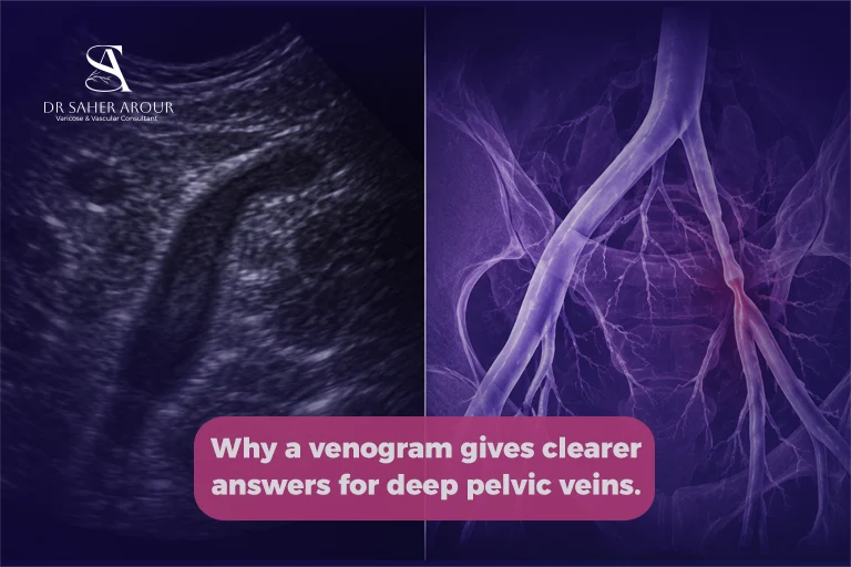
Conventional contrast venography is a reference standard test for DVT diagnosis [2]. When we review the images, a blood clot is clearly identified as an intraluminal filling defect or the nonfilling of a venous segment with a sharp cutoff [3]. However, modern contrast venography is seldom performed just to take diagnostic pictures; we use it primarily when concurrent intervention is planned to actively treat the problem we find [1].
When Does a Vascular Surgeon Order a Venogram Instead of an Ultrasound?
We always prefer to start with the least invasive option. Compression ultrasonography (CUS) is the first-line imaging test for a suspected lower-extremity DVT [4]. Clinical guidelines favor ultrasound over venography for the initial diagnosis of a first lower-extremity DVT [5], and the American Society of Hematology confirms that extremity DVT diagnosis primarily uses ultrasonography [6]. We use it widely because it is noninvasive, easily available, repeatable, and highly accurate for detecting symptomatic DVT [7].
However, ultrasound has structural limits. When those limits prevent us from safely diagnosing your condition, a venogram becomes essential.
Situation 1: Ultrasound Cannot Image the Pelvic or Abdominal Veins Adequately
One major drawback is that ultrasound has limited visualization within the pelvis [7]. In most patients with suspected May-Thurner syndrome, the deep veins in the pelvis cannot be fully visualized by ultrasound [8]. In these cases, a stenosis greater than 50% of the venous luminal diameter indicates left common iliac vein compression [9].
If iliofemoral or iliocaval involvement is suspected, alternative techniques like CT venography (CTV) or MR venography (MRV) are often required for a detailed anatomical assessment [10]. Contrast venography can effectively image pelvic veins and the inferior vena cava (IVC) when they are poorly displayed by other techniques [2]. CTV is also excellent for evaluating pelvic veins and the IVC that are difficult on ultrasound [1].
Situation 2: Planning Complex Catheter-Based Treatment
Contrast venography is strictly used when concurrent interventions are planned [11]. In these scenarios, same-session endovascular treatments like mechanical thrombectomy, intravascular ultrasound (IVUS), angioplasty, and stenting can be performed with 100% technical success [12].
Venous stenting is highly reliable. A large meta-analysis of 5,154 patients showed venous stenting technical success was 97% to 100%, with zero periprocedural deaths [13]. For acute DVT, 1-year primary patency after stenting was 91%, with cumulative patency at 97% [13]. In one specific iliofemoral DVT and May-Thurner cohort, the 12-month primary patency after thrombectomy and stenting was 73.5% for inpatients and 86.7% for outpatients [12].
To ensure this long-term success, precise measurements are critical. A residual venous stenosis greater than a 50% diameter reduction, or a pressure gradient greater than 2 mmHg, is a commonly used threshold for considering stenting [14]. Leaving a residual venous stenosis of 50% or more following a thrombectomy predicts reduced long-term patency [15]. Therefore, adjunctive stents are frequently required after thrombus removal to treat an uncovered lesion or significant residual thrombus [16]. During these precise interventions, IVUS has widely replaced older imaging methods because it boasts a higher sensitivity of over 98% [12]. IVUS directly visualizes luminal stenosis and vessel wall compression much more accurately than conventional venography [10].
Situation 3: When Previous Tests Have Given Conflicting Results
There are times when your physical symptoms and your ultrasound report simply do not match up. We use venography when noninvasive imaging is equivocal [2]. As a patient, this uncertainty can be incredibly frustrating, but we have precise tools to find the exact answer. Standard venography can confirm a DVT diagnosis when ultrasonography findings are equivocal [2]. When duplex scanning is inadequate, venography or CT venography should be considered [2]. CT and MRI also act as excellent problem-solving tools to visualize thrombosis in central veins when initial tests leave us unsure [1].
Types of Venography: Which One Is Used for Which Condition?
We choose the specific type of venogram based on the anatomical details we need to see and your overall medical safety.
| Imaging Modality | Primary Advantage | Clinical Application | Main Limitation |
| Conventional Venography | Real-time visualization and immediate treatment. | Used when concurrent endovascular interventions (like stenting) are planned [11]. | Invasive; requires ionizing radiation and iodinated contrast [1]. |
| CT Venography (CTV) | Accurate thrombus detection and quick acquisition. | Excellent for evaluating pelvic veins and the IVC that are difficult on ultrasound [1]. | Exposes the patient to ionizing radiation and contrast dye [11]. |
| MR Venography (MRV) | Highly detailed soft tissue and perivascular imaging. | Clarifies surrounding pathology with zero ionizing radiation [11]. | Can take longer; non-contrast options exist to avoid dye entirely [2]. |
When comparing CT venography vs ultrasound, CTV gives us a much broader cross-sectional view. On a CTV scan, a thrombosed vein appears clearly as a dark filling defect [17].
MR venography provides excellent anatomical detail and accurate thrombus detection without any ionizing radiation [11]. MRV is incredibly accurate; MRV for ilio-femoro-caval DVT showed a sensitivity up to 0.98 for occlusion [11], with strong interobserver agreement (kappa value 0.91) [11]. Furthermore, MRV is a sensitive pelvic-vein DVT test that completely avoids radiation and contrast [11]. Non-contrast MR venography successfully avoids ionizing radiation, vein cannulation, and contrast injection [2]. In one prospective study, non-contrast MRV achieved 100% sensitivity and 99.6% to 100% specificity for diagnosing a thrombus [18]. Similarly, black-blood thrombus MRI for acute DVT achieved 95.2% sensitivity, 98.6% specificity, and 97.7% accuracy [19].
What Happens During a Catheter Venogram: Step by Step
Knowing exactly what to expect helps reduce the anxiety many patients feel before a vascular procedure. We always begin by cleaning and numbing the access site, usually on your leg or groin, so you remain comfortable.
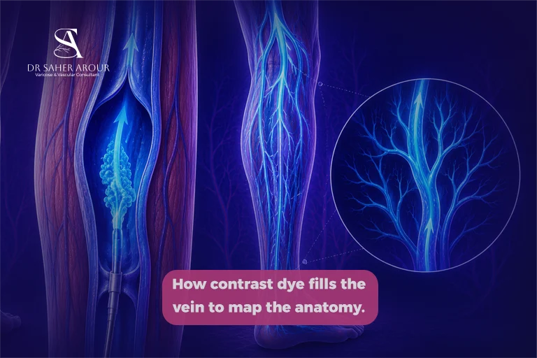
A tiny, flexible catheter is gently guided into the targeted vein. Contrast dye is then slowly injected, and we ask you to hold still while the fluoroscopy camera takes real-time pictures. If we locate a significant narrowing, we can immediately shift from diagnosing the issue to fixing it, performing interventions smoothly in the same setting. Finally, the catheter is removed, and we hold light pressure on the area to stop any bleeding.
What the Venogram Results Mean: Reading the Key Findings
Normal Venogram: What It Shows
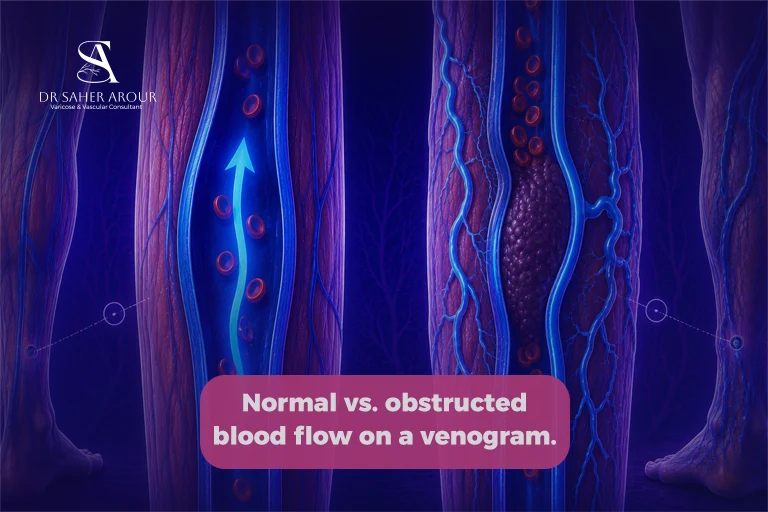
A normal result means the contrast dye travels freely through your veins. We look for a smooth, unbroken flow of dye, which confirms your vein is patent and open, without any strictures or clots slowing the blood down.
Abnormal Findings and What They Indicate
If there is an active clot or blockage, the imaging will pinpoint it immediately. Here is exactly what we look for:
- Filling defects: We observe an intraluminal filling defect or a nonfilling segment with a sharp cutoff, signaling a clot [3].
- Collateral veins: Acute thrombosis does not typically present with significant alternative venous drainage [10]. The absence of collateral circulation suggests an acute DVT, while their presence points to a chronic DVT [3].
- Residual stenosis: We measure the severity of any narrowing to see if it reaches the greater than 50% stenosis threshold that often requires a stent to keep the vein open safely [14].
Is a Venogram Painful or Risky?
We take every precaution to ensure your safety. Conventional venography does carry patient radiation exposure and the possibility of adverse reactions to the iodinated contrast medium [3]. Digital subtraction angiography and venography share these exact risks, including radiation exposure and allergic or nephrotoxic effects of iodinated contrast agents [17]. There is also a small risk of contrast reaction and iatrogenic venous thrombosis [1]—which simply means a blood clot that forms as a direct result of the catheter irritating the vein.
Thankfully, most iodinated contrast reactions are mild chemotoxic symptoms, though rare, unpredictable, and life-threatening hypersensitivity can occur [20]. The most significant risk factor for this is a history of iodinated contrast hypersensitivity [20], which is why we review your medical history carefully beforehand.
If we proceed with a catheter-based treatment, bleeding is a factor we manage carefully. In the major stenting meta-analysis, minor bleeding was reported in up to 5% of patients, and major bleeding occurred in only 0.5% upon intervention [13]. Your comfort and safety remain our absolute priority throughout the entire process.
FAQ Section: People Also Ask
How is a venogram different from an ultrasound?
A venogram uses contrast dye and X-ray imaging to directly visualize blood flow inside veins, while ultrasound uses sound waves and is non-invasive. Venography is typically used when ultrasound is inconclusive or cannot adequately image certain veins, especially in the pelvis or abdomen.
Does a venogram hurt?
Most patients experience only mild discomfort during a venogram. We use local anesthesia, but you may feel a brief warm sensation when the contrast dye is injected. Because the procedure involves a needle and catheter, some minor pressure at the insertion site can occur.
How long does a venogram take?
The duration of a venogram varies widely based on what we find. Since modern contrast venography is seldom performed unless a concurrent intervention is planned, complex interventional cases will naturally take more time than a simple diagnostic scan.
Do I need to fast before a venogram?
In many cases, you may be asked to avoid eating or drinking for several hours before the procedure, especially since iodinated contrast dye is used. Your doctor will give specific instructions based on your medical condition and the type of venography planned.
Can a venogram be used to treat a vein problem in the same session?
Yes, in many cases a venogram is performed as part of an interventional procedure. If a blockage or narrowing is identified, treatments such as angioplasty, stenting, or thrombolysis are often safely performed during the same session.
References
[1] Contrast venography: state of the art
[2] The diagnosis of deep vein thrombosis
[3] Conventional X-ray venographic criteria of DVT
[4] Ultrasound is the first-line imaging test for suspected lower-extremity DVT
[6] American Society of Hematology 2018 guidelines for management of venous thromboembolism
[7] Venous ultrasonography (US) is most widely used for the initial assessment of DVT
[8] Pelvic deep veins cannot be fully visualized by ultrasound
[9] Stenosis greater than 50% of the venous luminal diameter
[10] Alternative imaging techniques such as CTV or MRV
[11] The advantages of CTV and MRV
[12] Same-session endovascular treatment technical success
[13] Venous stenting technical success and safety
[14] Significant stenoses (>50% diameter reduction)
[15] Residual venous stenosis ≥50% predicts reduced long-term patency
[16] Adjunctive stents are frequently required
[17] Digital subtraction angiography/venography risks
[18] Non-contrast MR venography for DVT